The OASIS project for the episiotomy planning
In 2014, an interesting study on the use of multichannel surface EMG for studying the anal sphincter in pregnant women was published. The European OASIS (Obstetric Anal Sphincter Injuries) project was coordinated by Professor Roberto Merletti and involved nine hospitals from five different European countries, which included 511 women in the study.
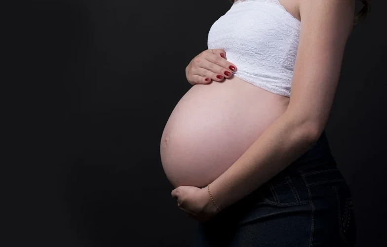
Incontinence in women after delivery
The pelvic floor is a diaphragm that supports the abdominal muscles, particularly the urogenital ones: a dysfunction of the muscles that make it up can lead to incontinence. This condition still involves a social stigma and leads to a reduced quality of life for patients.
In particular, the fecal incontinence on which the study focused is partly caused by a deficit of the external anal sphincter muscle (EAS). The remarkable fact is that it seems to occur particularly after delivery, with a prevalence of between 2% and 19%!
But what is the reason for the deficit?
The episiotomy technique
Recent studies have shown that iatrogenic asymmetries of the pudendal nerve innervating the EAS may contribute to incontinence symptoms: episiotomy (the surgical widening of the vaginal canal during labour to reduce the risk of a complex tear due to pressure from the baby’s head) is one of them.
Unlike skeletal muscles, EAS seems to have a distribution of innervation zones (IZs) that is totally random and cannot be determined by theory alone: if these zones are injured by the episiotomy, the sphincter loses part of its voluntary control and this could lead to incontinence.
However, did you know that there is no general indication regarding the optimal position where to make the incision? Despite that, most of the time this technique is performed laterally to the right just because the gynaecologist is right-handed.
It is reasonable to think that making the incision in the portion with less IZs can reduce the risk of denervation of the muscle.
But how do we identify this area?
The added value of surface EMG: applications in pregnancy for episiotomy
An electromyographic analysis of the anal sphincter was conducted: in the weeks prior to birth, IZs were identified using a cylindrical probe with 16 circumferential electrodes, and then re-evaluated two months after childbirth.
Neither caesarean section nor natural childbirth changed the number of IZs, even in women who had a spontaneous laceration. In contrast, many women who had undergone an episiotomy showed partial denervation of the EAS. Future studies are needed to confirm that faecal incontinence is related to EAS denervation.
Meanwhile, performing an EMG screening in the weeks leading up to delivery would make it possible to know the areas with the highest density of IZ and to act accordingly by not performing an eventual episiotomy there. This could also involve changing the surgical procedures or develop new instrumentation to perform the cut on the left, if needed.
If you still want to know more, this volume is right up your alley!

