
The importance of paying attention to the electrode placement
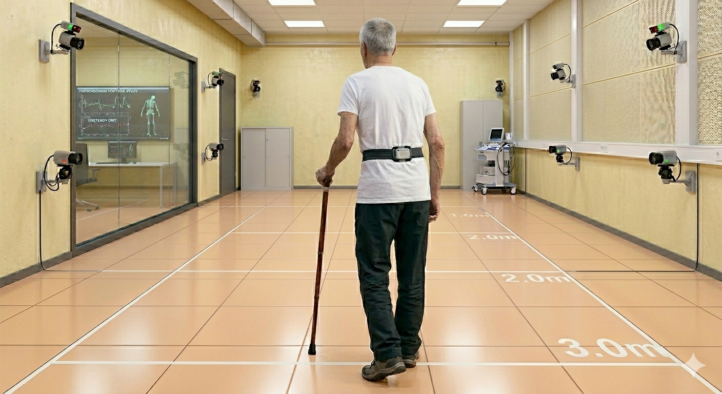
When applying an electrode for surface electromyography (sEMG), we must pay attention to its correct placement, because signal detection can be influenced by several factors and lead to erroneous conclusions during evaluation.
For this purpose, specific protocols were developed, like the SENIAM (Surface ElectroMyoGraphy for the Non-Invasive Assessment of Muscles) one.
But, in everyday clinical practice, how much can we get wrong in positioning the electrodes without altering the signal and, consequently, the interpretation of the results?
Our study
In 2007, the Movement Analysis Laboratory of Correggio (Azienda USL – IRCCS of Reggio Emilia), directed by dr Isabella Campanini PT, Ph.D., conducted a study concerning this point in collaboration with the LISIN of the PoliTO coordinated by Dr. Roberto Merletti and Prof. Dario Farina at the University of Aalborg, Denmark.
Although a few years have passed since its publication, the message is still relevant, given the increased use of this method in several hospital research laboratories.
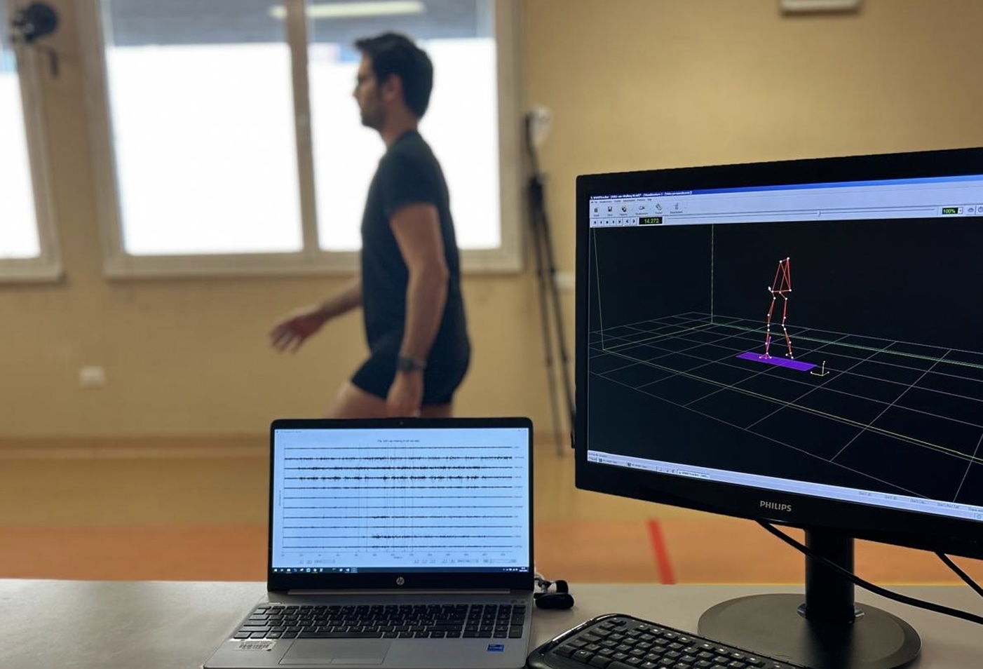
Ten healthy subjects were recruited to analyze the EMG envelope of some leg muscles during the gait cycle, through multiple electrode locations. It was hypothesized that the different placement of electrodes on the same muscles could lead to different characteristics of EMG activity.

Reproduced with the permission of Dr. Campanini and Colleagues from Campanini et al. 2007 on Journal of Electromyography and Kinesiology
A grid of 4 x 3 electrodes was placed on the tibialis anterior (TA), peroneus longus (PL), gastrocnemius medialis (GM), and gastrocnemius lateralis (GL), while a grid of 3 x 3 was placed on the soleus (SO). Each muscle was assessed individually during walk at self-selected speed. The basographic signal was also recorded using a foot-switches to determine heel strike and toe off instants.
The results and the recommendations
As shown in the figure below, TA electrodes must be placed near the tibia bone. Electrodes on PL must be placed carefully to prevent crosstalk from the nearby dorsiflexors (e.g., Extensor digitorum longus).
GM and GL signals were substantially unaffected by small changes in electrode placement. Instead, SO electrodes must be placed correctly according to the SENIAM recommendations (medially and below, not centrally); otherwise, the variability ratio of the signal increase, leading to a misinterpretation of the results.
The integral article can be found here.
The study underlined the complexity of determining the intensity of muscular activity during gait using sEMG, recommending always checking for possible crosstalk between muscles after electrode placement.

Reproduced with the permission of Dr. Campanini and Colleagues from Campanini et al. 2007 on Journal of Electromyography and Kinesiology

Reproduced with the permission of Dr. Campanini and Colleagues from Campanini et al. 2007 on Journal of Electromyography and Kinesiology
