The equinus foot deformity
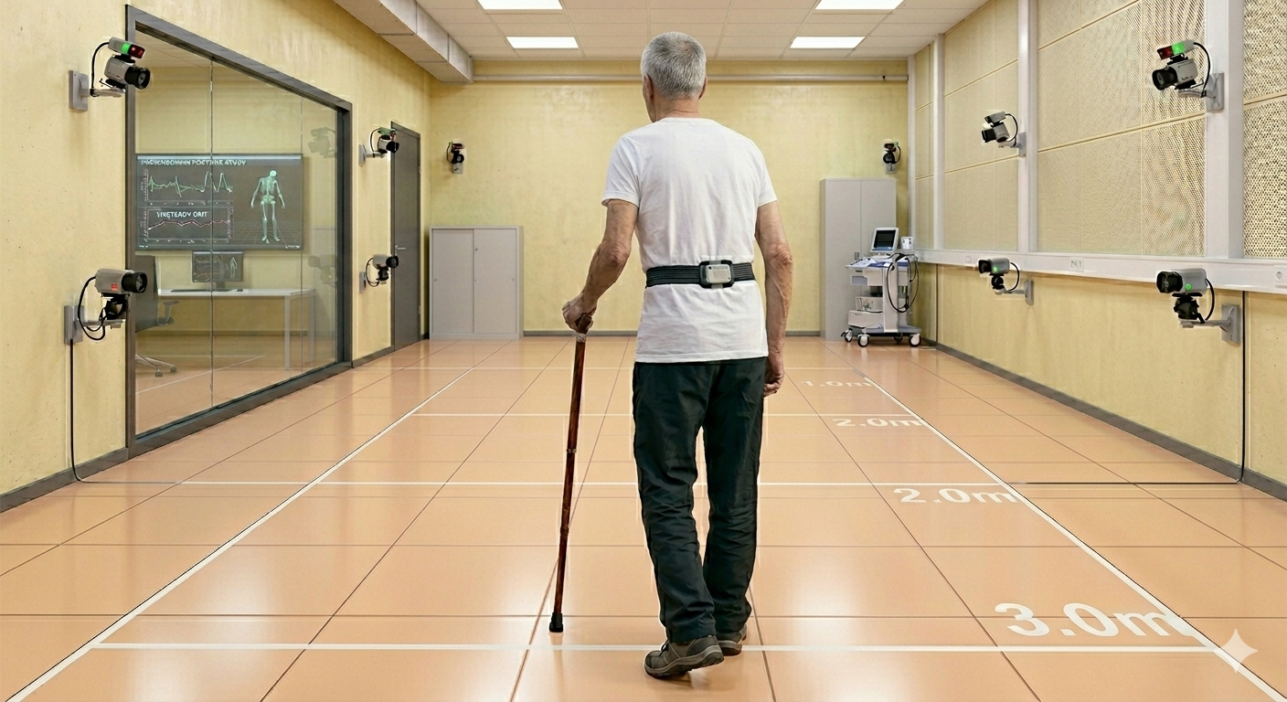
Equinovarus foot deformity is the most frequent lower limb acquired deformity after a stroke. It can be caused by several causes including central and peripheral phenomena involving agonists and antagonistic ankle muscles. The supination deviation may as well be due to the imbalance between Tibialis Anterior and Extensor Digitorum Longus (EDL), respectively the main medial and lateral dorsiflexor muscles, when calf muscles act as a posterior brake.
Despite his fundamental role is clearly defined in books and manuals, EDL is hardly ever considered in clinical practice and basically never mentioned in the literature about equinus foot deformity. We therefore created a biomechanical musculoskeletal model to measure the individual role of dorsiflexor and plantarflexor muscles in the manifestation of the equinus foot deformity.
New collaborations
This was a great opportunity to work with respectable bioengineers from the Politecnico di Milano, dr. Carlo Frigo, and the Italian Council of National Research of Lecco, dr. Cristina Brambilla. Of course, the clinical experience of the colleague physiotherapist dr. Davide Mazzoli has been an added value to the work.
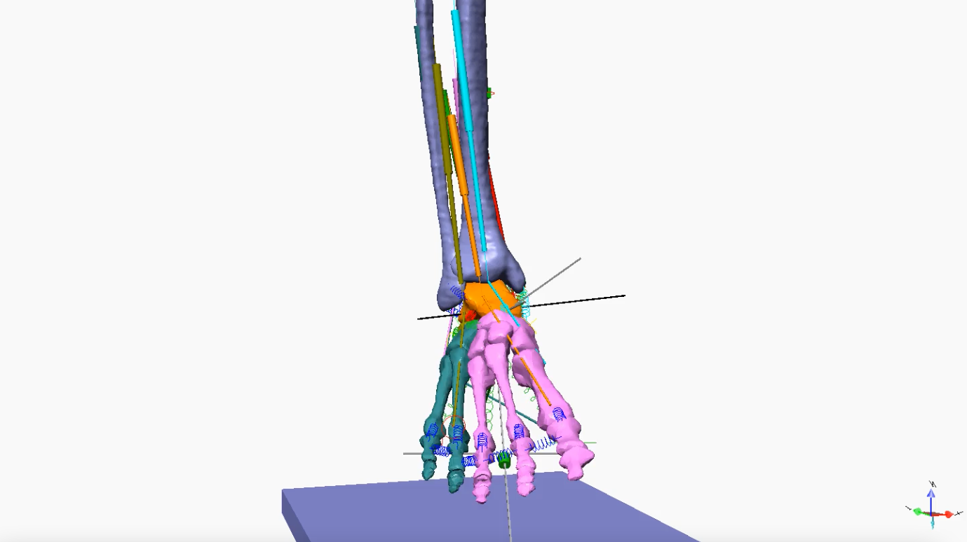
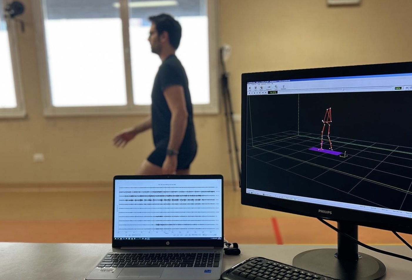
Our model analyzing the role of TA and EDL
In the model, we simulated different combinations of muscle contractions and movement constraints, reproducing what usually happens in stroke patients’ triceps surae, which usually go toward progressive contracture and shortening.
We found that neither Tibialis Anterior nor EDL alone can produce a satisfying dorsiflexion, rather causing a foot deviation on the frontal plane (i.e., supination and pronation, respectively). Only their synergistic action can lead the foot to dorsiflexion suitable for most daily life activities.

The paper aimed to increase clinicians’ and researchers’ awareness of the role of EDL, usually neglected for the Tibialis Anterior which is mistakenly deemed as the main dorsiflexor muscle. Moreover, we demonstrated how triceps surae contractures can influence the onset of frontal foot deviations, suggesting early treatment options to be considered.
The paper is open-access and can be downloaded here.
Extensor Digitorum Longus contraction alone
Tibialis Anterior contraction alone
Extensor Digitorum Longus and Tibialis Anterior combined contraction
Being a bioengineer in rehabilitation is a tremendous opportunity to collaborate with experts in many fields, learn new things, and develop ideas and projects that will serve the patient.
Contact us through the form if you want to collaborate with us.
